cat dental chart roots
And hyperplasia mobility furcation involvement and other oral pathology can all be recorded on a dental chart. Permanent teeth usually begin to appear at around 4 to 7 months see Table.

World Small Animal Veterinary Association Global Dental Guidelines Niemiec 2020 Journal Of Small Animal Practice Wiley Online Library
Root Canal SingleDoubleTriple Restorations Crown Cap Crown Impression Crown Lengthening Crown Preparation BondingRestoration _____ Glass Ionomer _____ Exodontics Alveoloplasty Crown Reduction.
. The cause of tooth resorption is not known. In those teeth with two or more roots the point where they diverge is called the furcation angle. The following images.
Probing depths up to 6 points per tooth of gingival recession. The dental formulae are as follows. Missing rotated and fractured teeth.
This can be a bifurcation or a trifurcation. I 33 C 11 P 32 M 11. Over time all areas of an affected tooth from root to crown may become involved.
Effective feline dental work cannot be done without general anesthesia. All findings should be recorded on a dental chart. Incidence reports list a range from 30 to 60 for cats affected by this oral condition.
Milinda Lommer DVM Created Date. The mesial surface of the first incisor is next to the median plane. Mesial and distal.
Title cat dental chart simplified Author. Tooth resorption is a process by which the dentin in a single toothor several teeth simultaneouslyerodes and eventually becomes irreparably destroyed. 26 Lateral incisor.
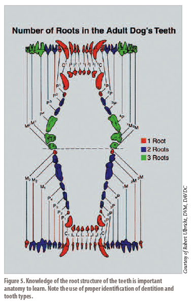
In man the design of the teeth is a reflection of eating habits as humans tend to be meat eaters so teeth are formed for cutting tearing and grinding food. Cats are obligate carnivores although pet cats consume a lot of plant material if they are fed dry cat food. One root for first tooth and number 11 305 311 and 405 411 All others - two roots.
28 First Premolar1st Bicuspid. Mandibular 1st molar cat ends in 09 ie. Look for facial asymmetries wounds and ability to properly close the mouth crepitus and bite.
Like us humans they will have 2 sets of teeth. I 33 C 11 P 44 M 23. Feline Dental Assessment Chart Author.
I 33 c 11 p 33. Mesial and distal are labeled in Photos 2A and 2B. Root Teeth may have one or more roots.
The human teeth dental chart illustrates the types and working surfaces of the four classes of teeth. At the end of the root is the apex which can have a single foramen humans a multiple canal delta arrangement cats and dogs or rema in open as in herbivores. Enamel is essentially impervious preventing bacteria from entering into the tooth.
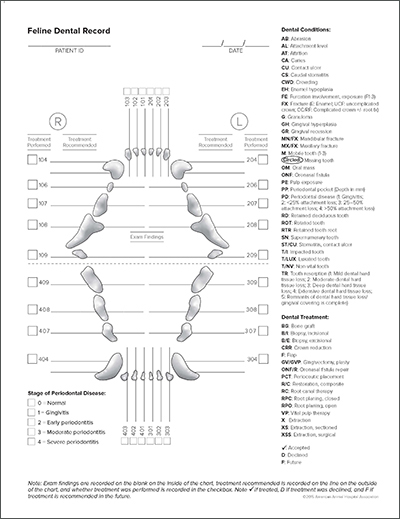
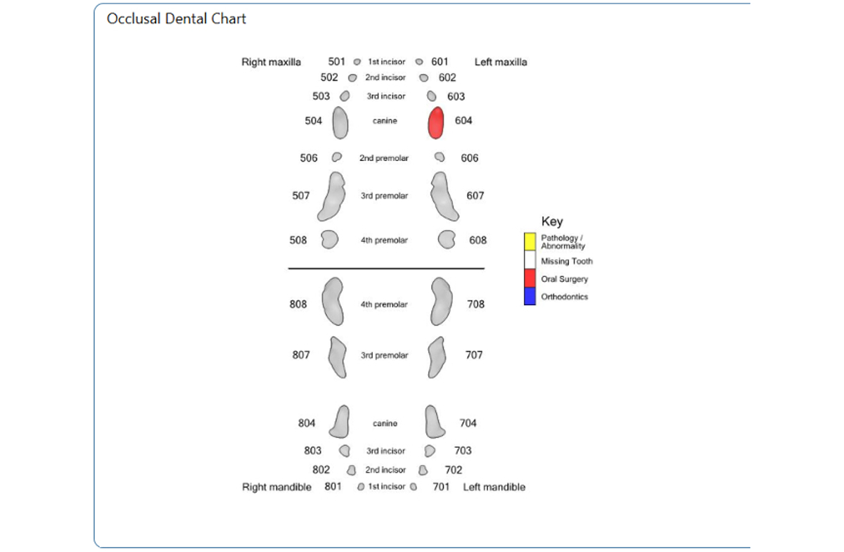
Beneath the enamel is a different dental hard tissue known as dentin. Circle missing teeth lateral view Fracturedabraded ABattrition AT teeth note to right with abbreviation shade in abnormality note abbreviation Discolored teeth shade in and note color adjacent Resorptive lesions shade in resorptive lesions on tooth at lateral view at topbottom and indicate stage of resorption. Dental Anatomy of Cats.
I 33 c 11 p 32. Feline TR is a very common problem. 29 Second Premolar2nd Bicuspid.
2I3I3 C1C1 P3P2 M1M1 30 teeth. None of the teeth of cats including their molars have grinding surfaces. Tooth resorption is the most common cause of tooth loss in cats and between 30 and 70 of cats show some sign of this destructive process.
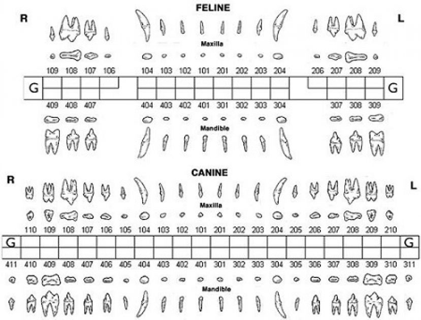
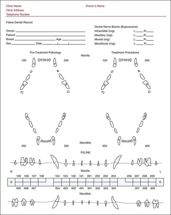
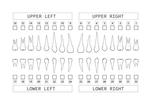
This basic tooth chart can act as your guide in tracking your treatment and knowing which tooth or quadrant is being worked upon or is complete. In the cat all the incisors and canine teeth have 1 root the maxillary 2nd premolar has 1 root the 3rd premolar has 2 roots and the 4th premolar has 3 roots while the. In the cat all the incisors and canine teeth have 1 root the maxillary 2nd premolar has 1 root the 3rd premolar has 2 roots and the 4th premolar has 3 roots.
Most cats have 26 deciduous teeth and 30 permanent teeth. 12 Risk factors include increasing age and the presence of other dental disease including additional TR lesions. Normal Period Depth Cats.
Tartar on crown and root. Normal Perio Depth Dogs up to 3mm. In the cat all the incisors and canine teeth have 1 root the maxillary 2nd premolar has 1 root the 3rd premolar has 2 roots and the 4th premolar has 3 roots while the maxillary 1st molar has 2 roots.
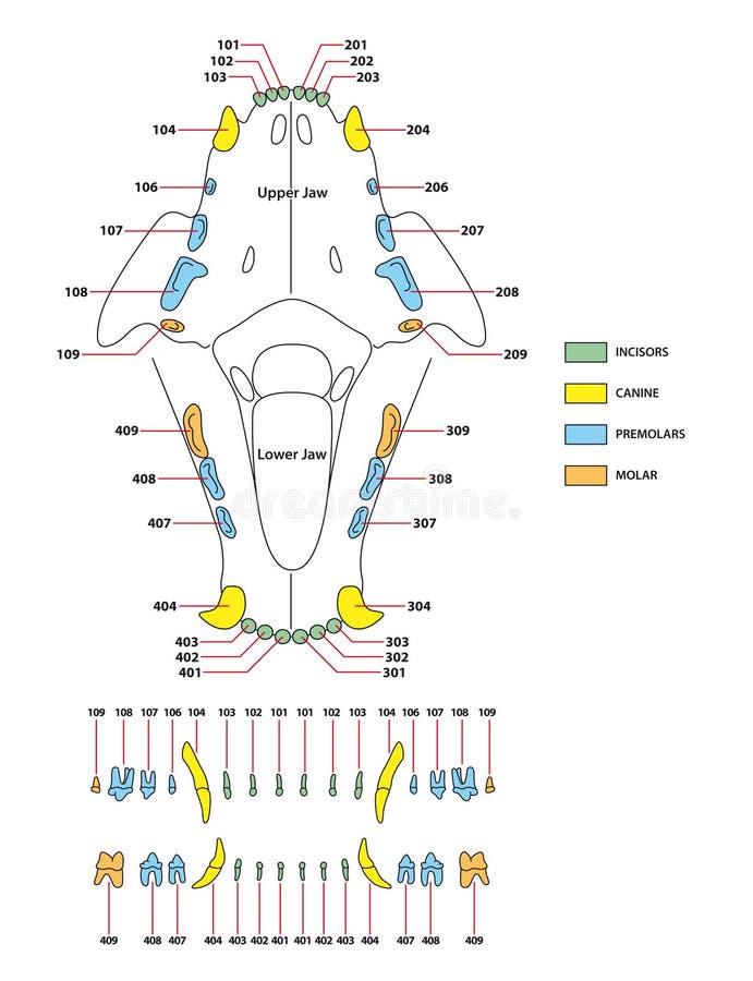
There are several reasons for this and they all come back to one fact. 3 Dental radiographs are required for proper diagnosis and treatmentFeline tooth resorption TR a common disease in cats characterized. One tooth with one root 105 and 205 Two teeth with two roots 106 107 and 206 207 Three teeth with three roots 108 109 110 and 208 209 210 Memory device for normal root numbers for canine permanent caudal mandibular teeth in each quadrant.
Periodontal disease in which the support structures of the toothcementum periodontal tissue ligament. Tooth resorption is a process in which the tooth structure breaks down beginning inside the tooth and often progressing to other parts of the tooth. The distal surface is opposite from the mesial surface.
On other teeth it is the surface directed toward the first incisor the surface adjacent to the tooth in front of it. 32 Third Molar Wisdom tooth. A retained deciduous canine in a cat.
Maxillary PM4 cat ends in 08. FELINE DENTAL EXAMINATION 2009 Veterinary Learning Systems BE Biopsy excisional BI Biopsy incisional CA Caries CI Calculus index 1 2 3 CR Crown restoration FI Furcation index 1 2 3 FX Fractured tooth or jaw G Granuloma GI Gingival index 1 2 3 GH Gingival hyperplasia GR Gingival recession MI Mobility index 1 2 3 OM Oral mass PC Pulp capping PE Pulp exposed. Education of colleagues and clients.
The permanent dental formula for adult cats is. Dogs also are carnivores but do have grinding surfaces on their molar teeth. Dentin contains approximately 300-400000 small.
The crown of a health tooth is covered by enamel. Prior to anesthesia the head should be assessed. They are used for plenty of normal tasks and it.
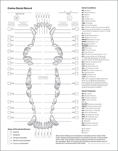
A tooth root abscess develops when bacteria enter the exposed root canal of the tooth. AVDS Canine Dental Record 2. The deciduous incisors begin to erupt at 2 to 4 weeks of age and the deciduous premolars at 5 to 6 weeks of age.
Feline dental chart A cats dental chart is a permanent record of their health that includes any abnormalities the skull type and occlusion findings. IM3 have kindly agreed to share the Australian Veterinary Dental Society Dental Charts for dogs cats rabbits guinea pigs. This disease differs from.
They clearly evolved to eat meat. Cat Dental Costs If your cat should need dental work and most will eventually you will no doubt see a bill that is substantially higher than what you get from your own dentist when you go for your regular dental cleaning. A tooth is a hard bony appendage that develops on the jaw to pulverize food.
0 - 05.
2
Dental Charts

Cat S Tooth Resorption Case Vets On The Balkans An Online Journal For Veterinarians From The Balkans

74 Canine Dental Chart Illustrations Clip Art Istock
Screenshots Animal Dental Chart

Dental And Oral Cavity Veterian Key

Dentalabels Charting System Smartpractice Veterinary
Dental And Oral Cavity Veterian Key

How Many Teeth Do Cats Have Cat Veteran
Unconscious Oral Evaluation
Teeth Charting Images Browse 4 442 Stock Photos Vectors And Video Adobe Stock
2
Adult Dental Chart Stock Illustrations 172 Adult Dental Chart Stock Illustrations Vectors Clipart Dreamstime

Dental Building Blocks Anatomy Charting And Cleaning Proceedings

Dentalabels Charting System Smartpractice Veterinary
Unconscious Oral Evaluation
Read This Dentistry Article By Jeanne Perrone Cvt

Cat S Dental Chart Animal Dentistry And Oral Surgery Specialists Llc Oshkosh Wi

Anhd 3170 Week 1 Dentistry Anatomy Of The Tooth Flashcards Quizlet